
Want fast and accurate ultrasound results?
Modern ultrasound technology has reached a level of precision where it can detect anomalies as small as 3–5 mm, significantly reducing the margin of diagnostic error.
By utilising advanced features such as Tissue Harmonic Imaging (THI) and AI-driven quality checks, Long Life Speciality Clinic, the best ultrasound scan centre in Mukundapur, ensures patients receive definitive reports, minimising the need for stressful, costly repeat scans.
In this blog, we’ll explore how modern ultrasound technology has advanced to deliver faster, more accurate results and help reduce errors and the need for repeat scans.
What Does “More Accurate Ultrasound” Mean?
When a doctor recommends a high-resolution scan, they aren’t just looking for a “prettier” picture.
Accuracy in sonography is the difference between a clear diagnosis and a vague “correlation required” report. For a patient, this means the technology can distinguish between a harmless fluid-filled cyst and a solid mass that requires immediate attention.
Better resolution, better contrast, and fewer artefacts
In the world of medical imaging, “resolution” refers to the machine’s ability to see two tiny structures as separate entities rather than one blurry blob. Modern machines at Long Life Speciality Clinic, the best ultrasound scan centre in Mukundapur, use high-frequency transducers that provide exquisite detail.
- Detail (Resolution): Allows the sonographer to visualise fine structures, such as fetal heart valves and tiny nerve bundles.
- Contrast (Separation): Helps differentiate between tissues of similar density, such as a tumour hiding within the liver.
- Artefact reduction: Modern software filters out “false pictures” or echoes that appear to be stones or shadows but aren’t actually there.
| Feature | Impact on Your Scan | Benefit to Patient |
| High Frequency | Superior detail in superficial organs | Accurate thyroid & breast staging |
| Low Frequency | Deeper penetration for abdominal scans | Clearer views of the liver and kidneys |
| Broadband Probes | Captures a wider range of data | Reduces the need for multiple probes |
Why fewer artefacts mean fewer “maybe” reports
Artefacts are visual “glitches” caused by physics, like sound waves bouncing off a rib or gas in the stomach. On older machines, these glitches often looked like real pathology, leading to “limited studies” in which the radiologist couldn’t be sure.
Advanced machines used at Long Life Speciality Clinic are designed to suppress these noise patterns, providing a “clean” image that leads to a definitive “Yes” or “No” rather than a “Maybe.”
What Is the Real Reason Ultrasound Sometimes Gets Repeated?
It is a common misconception that a repeat scan always implies a “bad” initial scan or a failing machine. Often, the human body is simply a complex environment that changes by the hour. Understanding why a second look is needed can bring immense relief to a family waiting for answers.
The 5 common causes of repeat scans
These are the causes for repeated scans
- Patient Factors: High BMI or excessive bowel gas can “mask” organs.
- Timing: A gallbladder scan is useless if the patient has just eaten, as the organ shrinks.
- Anatomy: Sometimes an organ is “hidden” behind the lungs or bone.
- Artefacts: Physical phenomena like “shadowing” from a rib.
- Operator Variability: A less experienced technician might miss a specific angle.
Note: A “Limited Study” note on your report usually means the doctor could see the organ, but not all of its borders clearly, due to gas or position. An “Abnormal Study” means they saw something, but need a specialised view (like Doppler) to confirm what it is.
Which Accuracy Upgrades Should You Look for in Modern Machines?
If you are looking for high resolution ultrasound in Kolkata or anywhere in India, you should know that not all machines are created equal. The “competitor-killer” features of modern systems are what truly protect you from misdiagnosis.
Tissue Harmonic Imaging (THI)
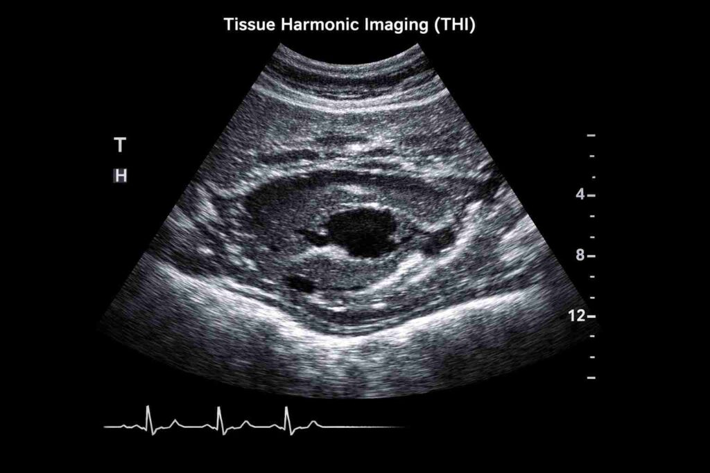
Tissue harmonic imaging, also known as native harmonic imaging, is a signal processing technique used in ultrasound. During the transmit phase of the pulse-echo cycle, an ultrasound beam insonates body tissues, generating harmonic waves due to nonlinear distortion. These harmonic waves are then utilised to enhance image quality and improve diagnostic accuracy. (SOURCE)
- Reduced Haze: Clears the “fog” in fluid-filled areas, such as the bladder.
- Sharper Borders: Makes the edges of organs or tumours stand out clearly.
- Deep Penetration: Maintains clarity even when looking at deep-seated structures.
Spatial Compounding and Speckle Reduction
Think of Spatial Compounding as taking photos from five different angles and merging them into one perfect shot. This eliminates “speckle”, the grainy texture that can hide small lesions. This technology is the gold standard for color doppler ultrasound accuracy, ensuring that blood flow is measured without “noise” interference.
Has Doppler Technology Become Smarter in Recent Years?
Doppler is the tool used to “see” and “hear” blood flow. In the past, “false blockages” were common if the patient moved slightly. Today, Doppler is incredibly sensitive.
Colour vs Power vs. Spectral Doppler
Each type serves a unique purpose in your diagnostic journey:
- Colour Doppler: Shows the direction and speed of blood (Red/Blue).
- Power Doppler: More sensitive; it detects even the tiniest blood flow in “slow” areas like small tumours, which Colour Doppler might miss.
- Spectral Doppler: Provides a graph of the flow, helping doctors calculate exactly how much a vessel is blocked.
This precision prevents “false alarms” about blockages and ensures that any missed flow issue is caught immediately.
Where Does 3D/4D Help—And Is It Just Marketing?
While 3D/4D ultrasound and advanced machine ads are everywhere, it’s important to know when they actually matter.
- Genuinely Helpful: 3D/4D is vital for detecting fetal facial anomalies (like cleft lip) or uterine abnormalities that a flat 2D image might miss.
- The Workhorse: For most abdominal or pelvic pain, the traditional 2D scan combined with Doppler remains the “gold standard” for accuracy.
How to Reduce the Chance of a Repeat Scan?
Expertise and technology can only do so much; your preparation is the final piece of the puzzle. Following these rules can increase your scan’s accuracy by up to 30%.
Preparation Rules That Matter
- Fasting (6–8 hours): Essential for abdominal scans to keep the gallbladder distended and bowel gas low.
- Full Bladder: For pelvic or early pregnancy scans, a full bladder acts as a “window,” pushing gas-filled intestines out of the way.
- Prior Reports: Always bring your old scans. Comparing a 2cm mass today to a 2cm mass from two years ago prevents an unnecessary “emergency” biopsy.
Revolutionising Diagnostics: The Power of Modern Ultrasound Technology
Modern ultrasound technology, featuring Tissue Harmonic Imaging and AI-guided protocols, has revolutionised diagnostic precision.
Don’t leave your health to chance with outdated technology. Are you ready to experience the clarity of a high-resolution scan? Book your precision ultrasound atLong Life Speciality Clinic, the best ultrasound scan centre in Mukundapur.
Frequently Asked Questions
Does a repeat scan mean something serious was found?
Not necessarily. Most repeats are due to “technical limitations,” such as bowel gas or the patient’s position, rather than a scary finding.
Can ultrasound be “wrong” even with the best machines?
Ultrasound is “operator-dependent.” The combination of expert sonographers and high-end machines delivers an accuracy rate above 95% for many conditions.
Should I get a second opinion if reports conflict?
Yes. If two reports from different centres disagree, seek a centre that uses high-resolution probes and has a senior radiologist review the raw footage.