In pregnancy, ‘visibility’ is the best form of prevention.
But did you know that different stages of your pregnancy require entirely different ultrasound technologies? A standard scan might confirm a heartbeat, but it won’t measure the blood flow to the placenta or the intricate chambers of a developing heart.
To bridge the gap between ‘watching’ and ‘diagnosing,’ we’re diving into the science of pregnancy-specific ultrasonography and why the right parameters make all the difference.
Let’s give you a brief idea about what pregnancy-specific ultrasonography is.
What Is Pregnancy-Specific Ultrasonography?
Pregnancy-specific ultrasonography, or obstetric ultrasound, is a safe, noninvasive imaging test using high-frequency sound waves to create real-time images of a developing fetus, placenta, and amniotic fluid within the uterus. It is used to confirm viability, determine gestational age, assess fetal growth, and screen for anomalies.
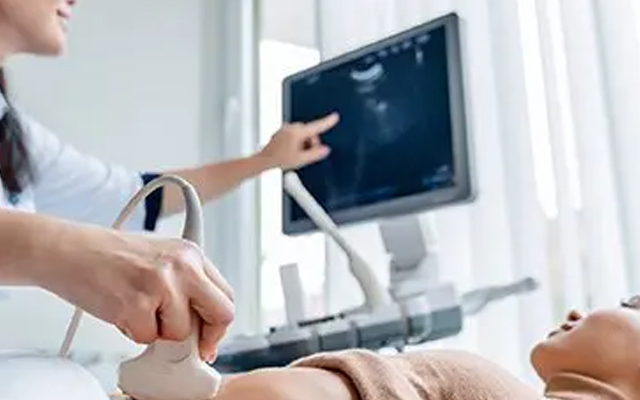
This is where obstetric ultrasound at Long Life Speciality Clinic in Mukundapur helps provide expectant mothers with reliable insights into the health and development of their pregnancy.
Before delving into a deeper understanding of the topic, a question: Did you have an idea of the different types of ultrasound scans? If not, let’s give you an idea about it.
When Pregnancy Ultrasound Is Recommended
Pregnancy ultrasounds are typically recommended at various stages of pregnancy to monitor the health of both the mother and the baby. Early ultrasounds, such as a pregnancy scan, are often performed to confirm the pregnancy, determine the due date, and check for any initial complications.
Types of Pregnancy Ultrasounds and What Each One Checks
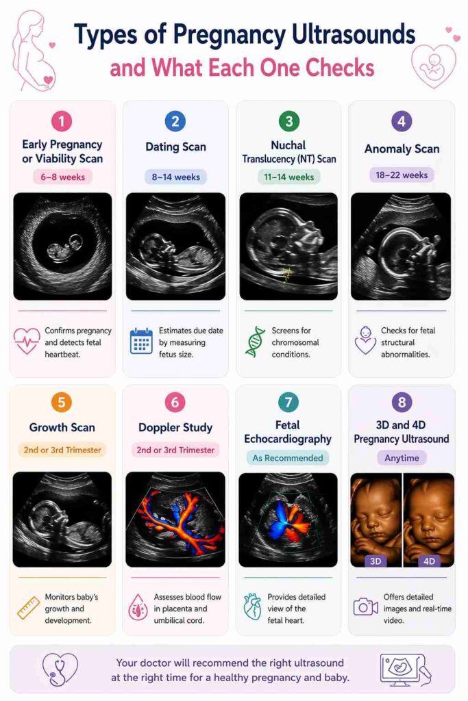
Pregnancy ultrasounds come in various types, each designed to monitor fetal health and development.
Early Pregnancy or Viability Scan
Typically performed between 6 and 8 weeks, this scan confirms pregnancy and detects the fetal heartbeat. It helps rule out complications such as ectopic pregnancies.
Dating Scan
Done between 8 and 14 weeks, the dating scan estimates the baby’s due date by measuring the fetus’s size and confirming the pregnancy’s progression.
Nuchal Translucency (NT) Scan
Between 11 and 14 weeks, the NT scan measures fluid at the back of the baby’s neck, screening for chromosomal conditions like Down syndrome.
Anomaly Scan
This scan, performed between 18 and 22 weeks, checks for fetal structural abnormalities, evaluating organs like the heart and brain to identify potential birth defects.
Growth Scan
Typically performed in the second or third trimester, this scan monitors the baby’s growth and development by measuring parameters such as head circumference and femoral length.
Doppler Study
A Doppler study assesses blood flow in the placenta and umbilical cord, crucial for the baby’s nutrient and oxygen supply.
Fetal Echocardiography
Recommended for potential heart defects, this specialised scan provides a detailed view of the fetal heart.
3D and 4D Pregnancy Ultrasound
Offering detailed images and real-time video, 3D and 4D ultrasounds help assess facial features and detect abnormalities that are not visible on 2D scans.
Dating Scan vs NT Scan vs Anomaly Scan vs Growth Scan
This table outlines their unique purposes, the best time to perform each scan, and what they can and cannot detect.
| Scan Type | Purpose | Best Time | What It Can & Cannot Detect |
| Dating Scan | Estimates the due date and confirms pregnancy viability | 8-14 weeks | Can determine gestational age; cannot detect birth defects |
| NT Scan | Assesses the risk of chromosomal abnormalities like Down syndrome | 11-14 weeks | Can detect risk of Down syndrome; cannot diagnose all chromosomal issues |
| Anomaly Scan | Checks for structural birth defects | 18-22 weeks | Can detect major organ and limb abnormalities; may miss minor issues |
| Growth Scan | Monitors fetal growth and estimates weight | 24 weeks onwards | Can detect growth restrictions; cannot identify structural abnormalities |
Are Pregnancy Ultrasounds Safe for Mother and Baby?
You’ve heard it a hundred times: Ultrasounds are safe, but you still wonder, “Is it really?”
As you see your little one on the screen, you realise just how reassuring it is to know that ultrasounds provide critical information without posing a risk to either you or your baby.
How to Prepare for a Pregnancy Ultrasound
The morning of your ultrasound, you’re feeling a mix of excitement and nervousness. You remember the instructions: drink plenty of water to ensure your bladder is full. You arrive at the clinic, ready for the scan.
With proper preparation, you’ll ensure a smooth, effective pregnancy ultrasonography test at Long Life Speciality Clinic in Mukundapur, providing valuable insights into your pregnancy’s progress.
Pregnancy is a vital part of a woman’s life, and you know about every stage:
First-Trimester Pregnancy Ultrasound Parameters
First-trimester ultrasound (roughly 5–13 weeks) evaluates pregnancy viability, dating, and morphology.
Gestational Sac and Yolk Sac
- The gestational sac appears around 5 weeks, confirming pregnancy.
- The yolk sac is a small, ring-like structure within the sac that supports early development.
Fetal Pole and Cardiac Activity
- The fetal pole appears by 5.5–6 weeks.
- Cardiac activity is typically detected at 6–7 weeks.
Crown-Rump Length (CRL)
- CRL measures the embryo from head to bottom, helping estimate gestational age.
Nuchal Translucency (NT)
- The NT scan (11–14 weeks) checks the fluid-filled space behind the fetus’s neck.
- It helps assess the risk for conditions like Down syndrome.
Nasal Bone and Early Screening Markers
- The nasal bone is assessed between 11 and 13+6 weeks.
- Its absence or underdevelopment is linked to chromosomal abnormalities like Down syndrome.
Second-Trimester Pregnancy Ultrasound Parameters
The second trimester (14–26 weeks) provides crucial information about fetal growth and development, helping to detect any potential abnormalities.
Biparietal Diameter (BPD)
BPD measures the width of the baby’s head and helps assess brain development and overall fetal growth.
Head Circumference (HC)
The HC measures the baby’s head circumference, which is important for tracking brain development and fetal growth.
Abdominal Circumference (AC)
AC measurement helps assess the size of the baby’s abdomen, providing valuable information on growth and nutritional status.
Femur Length (FL)
The length of the femur is measured to evaluate skeletal development and estimate the baby’s overall size and growth.
Detailed Fetal Anatomy Assessment
This scan evaluates the development of the baby’s organs and limbs, identifying any abnormalities in the brain, heart, spine, and other vital structures.
Placenta, Cervix, and Amniotic Fluid Evaluation
An assessment of the placenta’s position, cervix, and amniotic fluid levels helps ensure the pregnancy is progressing safely and healthily.
Third-Trimester Pregnancy Ultrasound Parameters
In the third trimester (27–40 weeks), the focus shifts to monitoring the baby’s growth and preparing for delivery.
Fetal Growth and Estimated Fetal Weight (EFW)
EFW is calculated to estimate the baby’s weight based on measurements of the head, abdomen, and femur, providing insights into fetal development.
Growth Trend and Interval Comparison
Comparing previous growth scans helps track whether the baby is growing at a healthy rate and alert doctors to any growth restrictions.
Amniotic Fluid Index (AFI) or Deepest Vertical Pocket
AFI measures the amount of amniotic fluid, which is important for fetal well-being. Low or high levels can indicate potential complications.
Fetal Position and Placental Location
This scan checks the position of the baby and the location of the placenta to assess if a C-section may be necessary.
Doppler Parameters for Fetal Well-Being
Doppler studies assess blood flow to the baby, ensuring they receive adequate oxygen and nutrients for proper growth.
When Additional or Repeat Pregnancy Scans May Be Needed
After a routine scan, your doctor mentions that a follow-up ultrasound may be needed to monitor your baby’s growth. You are surprised but reassured by the care being taken to ensure everything is going well. The repeat scan confirms that everything is progressing as expected.
While it wasn’t what you expected, you’re grateful that the medical team is thorough and proactive, giving you peace of mind that your baby’s health is a priority.
Let’s Recap
Pregnancy ultrasounds play a crucial role in monitoring both maternal and fetal health at different stages of pregnancy. From confirming pregnancy viability to checking for birth defects and tracking fetal growth, these scans offer invaluable insights.
If you’re looking for expert care, visit Long Life Speciality Clinic for a pregnancy ultrasound test in Mukundapur to get the best prenatal care today.
Call + 8334073407 / 9874807480
People Also Ask
Which scan is most important during pregnancy?
The anomaly scan is often considered the most important scan, as it checks for structural abnormalities in the baby. It is typically performed between 18 and 22 weeks and provides detailed insights into the fetus’s health.
What is the difference between a dating scan and an anomaly scan?
A dating scan helps determine the baby’s due date by measuring its size early in pregnancy, usually between 8 and 14 weeks. In Mukundapur, an anomaly scan is performed later (18-22 weeks) to check for birth defects and assess fetal development.
What does CRL mean in a pregnancy ultrasound?
CRL stands for Crown-Rump Length, which is the measurement from the top of the baby’s head (crown) to its bottom (rump). This measurement is used in early pregnancy ultrasounds to estimate gestational age and confirm the due date.
What are BPD, HC, AC, and FL in fetal biometry?
- BPD (Biparietal Diameter): Measures the width of the baby’s head.
- HC (Head Circumference): Assesses the circumference of the baby’s head.
- AC (Abdominal Circumference): Helps monitor fetal growth.
- FL (Femur Length): Measures the length of the baby’s thigh bone.
These measurements, taken during a pregnancy ultrasound in Mukundapur, help assess fetal development and growth.
Is a 3D or 4D ultrasound necessary in every pregnancy?
No, 3D and 4D ultrasounds are not necessary for every pregnancy. These advanced scans are typically used for specific purposes, such as assessing fetal features or providing a detailed view of the baby.